Publications & technical resources
Explore how DHO technology is facilitating scientific discovery

Thank you! Your submission has been received!
Oops! Something went wrong while submitting the form.

Three-dimensional localization precision of the double-helix point spread function versus astigmatism and biplane
Wide-field microscopy with a double-helix point spread function (DH-PSF) provides three-dimensional (3D) position information beyond the optical diffraction limit. We compare the theoretical localization precision for an unbiased estimator of the DH-PSF to that for 3D localization by astigmatic and biplane imaging using Fisher information analysis including pixelation and varying levels of background. The DH-PSF results in almost constant localization precision in all three dimensions for a 2 μm thick depth of field while astigmatism and biplane improve the axial localization precision over smaller axial ranges. For high signal-to-background ratio, the DH-PSF on average achieves better localization precision.

Performance limits on three-dimensional particle localization in photon-limited microscopy
We present the performance limits on three-dimensional (3D) localization accuracy of currently used methods of wide-field superlocalization microscopy. The three methods investigated are double-helix microscopy, astigmatic imaging, and biplane detection. In the shot-noise limit, Cramer–Rao lower bound calculations show that, among these techniques, the double-helix microscope exhibits the best axial and 3D localization accuracy over short as well as long depth-of-field systems. The fundamental advantage of engineered point-spread function systems, like the double-helix, stems from the additional degrees of freedom available to control diffraction in three dimensions over variable regions of interest.

In vivo three-dimensional superresolution fluorescence tracking using a double-helix point spread function
The point spread function (PSF) of a widefield fluorescence microscope is not suitable for three-dimensional superresolution imaging. We characterize the localization precision of a unique method for 3D superresolution imaging featuring a double-helix point spread function (DH-PSF). The DH-PSF is designed to have two lobes that rotate about their midpoint in any transverse plane as a function of the axial position of the emitter. In effect, the PSF appears as a double helix in three dimensions. By comparing the Cramer-Rao bound of the DH-PSF with the standard PSF as a function of the axial position, we show that the DH-PSF has a higher and more uniform localization precision than the standard PSF throughout a 2 μm depth of field. Comparisons between the DH-PSF and other methods for 3D superresolution are briefly discussed. We also illustrate the applicability of the DH-PSF for imaging weak emitters in biological systems by tracking the movement of quantum dots in glycerol and in live cells.

Localizing and Tracking Single Nanoscale Emitters in Three Dimensions with High Spatiotemporal Resolution Using a Double-Helix Point Spread Function
Three-dimensional nanoscale localization and tracking of dim single emitters can be obtained with a widefield fluorescence microscope exhibiting a double-helix point spread function (DH-PSF). We describe in detail how the localization precision quantitatively depends upon the number of photons detected and the z position of the nanoscale emitter, thereby showing a ∼10 nm localization capability along x, y, and z in the limit of weak emitters. Experimental measurements are compared to Fisher information calculations of the ultimate localization precision inherent in the DH-PSF. The DH-PSF, for the first time, is used to track single quantum dots in aqueous solution and a quantum dot-labeled structure inside a living cell in three dimensions.

Three-dimensional localization with nanometer accuracy using a detector-limited double-helix point spread function system
Accurate estimation of the three-dimensional (3D) position of particles is critical in applications like biological imaging, atom/particle-trapping, and nanomanufacturing. While it is well-known that localization accuracy better than the Rayleigh resolution limit is possible, it was recently shown that, for photon-limited cases, 3D point spread functions (PSFs) can be shaped to increase accuracies over a 3D volume [Pavani and Piestun, Opt. Express 16, 22048 (2008)]. Here, we show that in the detector-limited regime, the gain in accuracy occurs in all three dimensions throughout the axial range of interest. The PSF is shaped as a double helix, resulting in a system with fundamentally better 3D localization accuracies than standard PSF systems, capable of achieving single-image subnanometer accuracies.
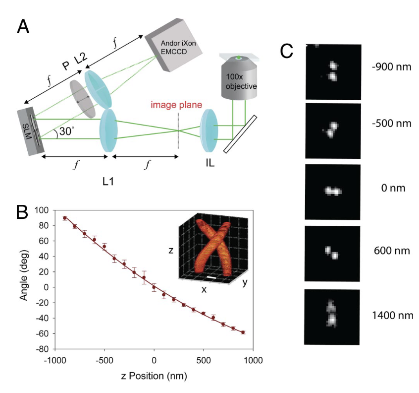
Three-dimensional, single-molecule fluorescence imaging beyond the diffraction limit by using a double-helix point spread function
We demonstrate single-molecule fluorescence imaging beyond the optical diffraction limit in 3 dimensions with a wide-field microscope that exhibits a double-helix point spread function (DH-PSF). The DH-PSF design features high and uniform Fisher information and has 2 dominant lobes in the image plane whose angular orientation rotates with the axial (z) position of the emitter. Single fluorescent molecules in a thick polymer sample are localized in single 500-ms acquisitions with 10- to 20-nm precision over a large depth of field (2 μm) by finding the center of the 2 DH-PSF lobes. By using a photoactivatable fluorophore, repeated imaging of sparse subsets with a DH-PSF microscope provides superresolution imaging of high concentrations of molecules in all 3 dimensions. The combination of optical PSF design and digital postprocessing with photoactivatable fluorophores opens up avenues for improving 3D imaging resolution beyond the Rayleigh diffraction limit.

Three dimensional tracking of fluorescent microparticles using a photon-limited double-helix response system
We demonstrate three-dimensional tracking of fluorescent microparticles, with a computational optical system whose point spread function (PSF) has been engineered to have two twisting lobes along the optical axis, generating a three-dimensional (3D) double-helix (DH) PSF. An information theoretical comparison in photon limited systems shows that the DH-PSF delivers higher Fisher information for 3D localization than the standard PSF. Hence, DH-PSF systems provide better position estimation accuracy. Experiments demonstrate average position estimation accuracies under 14nm and 37nm in the transverse and axial dimensions respectively. The system determines the 3D position of multiple particles with a single image and tracks them over time while providing their velocities.

High-efficiency rotating point spread functions
Rotating point spread functions (PSFs) present invariant features that continuously rotate with defocus and are important in diverse applications such as computational imaging and atom/particle trapping. However, their transfer function efficiency is typically very low. We generate highly efficient rotating PSFs by tailoring the range of invariant rotation to the specific application. The PSF design involves an optimization procedure that applies constraints in the Gauss-Laguerre modal plane, the spatial domain, and the Fourier domain. We observed over thirty times improvement in transfer function efficiency. Experiments with a phase-only spatial light modulator demonstrate the potential of high-efficiency rotating PSFs.

Depth from diffracted rotation
The accuracy of depth estimation based on defocus effects has been essentially limited by the depth of field of the imaging system. We show that depth estimation can be improved significantly relative to classical methods by exploiting three-dimensional diffraction effects. We formulate the problem by using information theory analysis and present, to the best of our knowledge, a new paradigm for depth estimation based on spatially rotating point-spread functions (PSFs). Such PSFs are fundamentally more sensitive to defocus thanks to their first-order axial variation. Our system acquires a frame by using a rotating PSF and jointly processes it with an image acquired by using a standard PSF to recover depth information. Analytical, numerical, and experimental evidence suggest that the approach is suitable for applications such as microscopy and machine vision.
No results found
Please try different keywords
Thank you! Your submission has been received!
Oops! Something went wrong while submitting the form.


No results found
Please try different keywords
Thank you! Your submission has been received!
Oops! Something went wrong while submitting the form.
No results found
Please try different keywords