High-resolution dynamic imaging of chromatin DNA communication using Oligo-LiveFISH

Three-dimensional genome dynamics play a critical role in cellular function and disease. However, real-time, live-cell DNA visualization remains challenging, as existing methods are often confined to repetitive regions, suffer from low resolution, or require complex genome engineering. This work by Zhu et al. introduces Oligo-LiveFISH, a high-resolution, reagent-based platform for dynamically tracking non-repetitive genomic loci in diverse cell types.
Why this is important
Aberrations in 3D genome architecture are linked to cancers, neurological disorders, and developmental abnormalities. However, 3D genome communication and its relationship with gene expression remain unclear due to the lack of robust approaches for real-time labeling and tracking non-repetitive genomic regions in live cells. Oligo-LiveFISH addresses this gap by offering a versatile platform for studying 3D genome dynamics and their links to cellular processes and disease.
The science
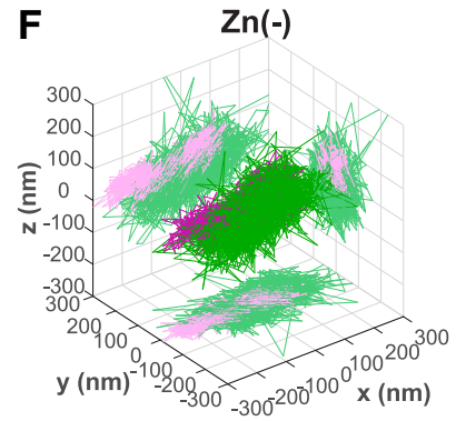
Oligo-LiveFISH uses a pool of fluorescent RNA guides to direct a DNA-binding protein called dCas9 to nearby sites along a chosen stretch of DNA. The labels’ combined signal makes even a non-repetitive region bright enough to track in living cells. Oligo-LiveFISH tracking and computational modeling revealed two distinct modes of chromatin communication. Additionally, studying FOS transcription under Zn stimulation showed slower dynamics between enhancer and promoter during active transcription.
How Double Helix Optics made it possible
The microscope used double-helix phase masks in its detection channels. Each mask shaped the target’s image into a point spread function with two lobes. The midpoint between the lobe centers provided x/y coordinates, and their calibrated orientations gave depth. Multicolor Oligo-LiveFISH imaging captured a 2 µm depth range in 3D with 20 nm spatial resolution at 50 ms per frame.